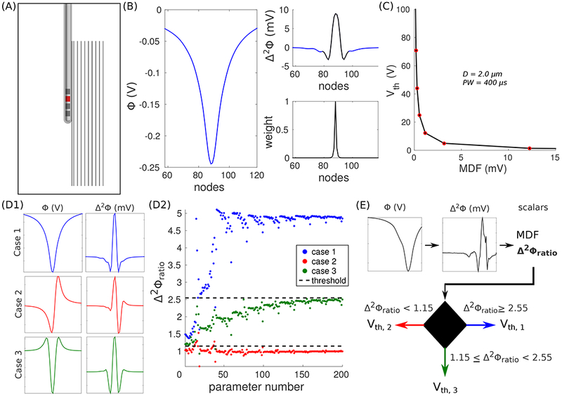
Figure 3.

DF-Howell. (A) Axisymmetric volume conductor model of Medtronic’s Model 3389 lead-in-a-box. Ten axons are oriented parallel to the long axis of the shaft. The lead is programmed in a monopolar cathodic configuration (C2−, red contact). The model is not drawn to scale. (B) Extracellular potentials (Φ, left) across an axon 1 mm from the surface of the lead. The modified driving force (MDF) is the inner dot product between the second difference of the potentials (Δ2Φ, top-right) and a set of weights (bottom-right). The black line in the top-right panel denotes where the weights are non-zero. (C) Activation thresholds (Vth) estimated with the models as a function of MDF. (D1) Φ and Δ2Φ for three different electrode configurations: Case 1 (C2− / IPG+, top), Case 2 (C2− / C3+, middle), and Case 3 (C2− / C1+, C3+, bottom). (D2) Δ2Φratio for all combinations of distance and fiber diameter. Δ2Φratio = Δ2Φmax / Δ2Φmin. The dashed lines denote decision boundaries that best separate the ratios between Cases 1 and 3, and Cases 2 and 3. (E) DF-Howell algorithm: Vth of a random axon in the cerebellothalamic tract (CbTT) is estimated using its MDF and Δ2Φratio.
