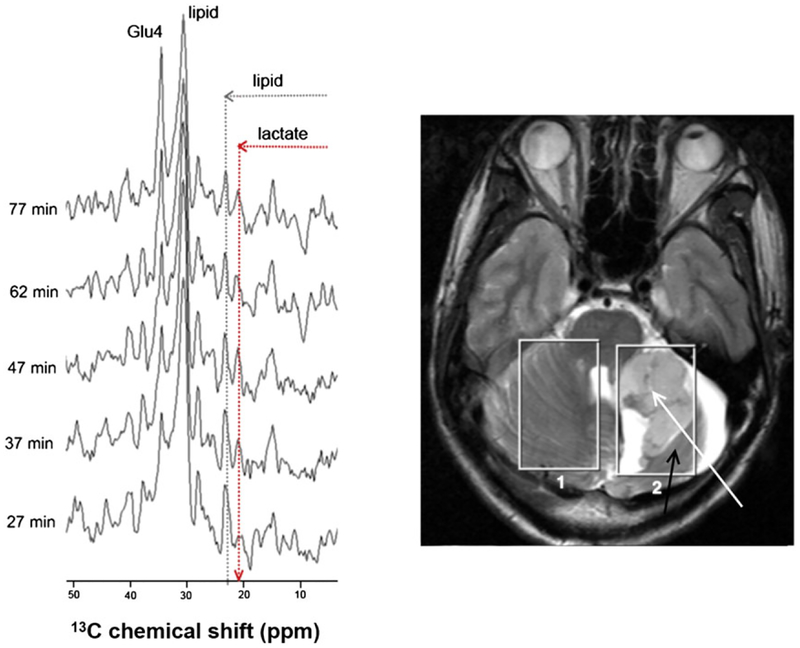
FIGURE 3.
13C MR spectra of the tumor region of a patient with high grade glioma after administration of [1-13C]-D-glucose (left). The 1H MR images (right) show the position of the two voxels (size 50 cm3) used for the 13C MRS. Voxel 1 is positioned in healthy cerebellum while Voxel 2 covers the tumor (white arrow) as well as some normal brain tissue (black arrow). The hyperintense areas are fluid filled. (Reproduced with permission from Reference 59)

