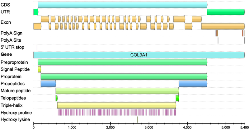
Fig. 1.

Structure of the human COL3A1 transcripts. The image shows the location of coding sequence (CDS), untranslated regions (UTR), 51 exons, polyA signals and sites, and the different functional domains. Locations of the proline residues that will be hydroxylated are also shown. The same color scheme was used in Figure 2. The annotations shown here differ slightly from those by Uniprot database (P02461–1), which are incomplete and did not incorporate domain expert knowledge. Also, there is no biological evidence that the Uniprot entry P02461–2 or the Ensembl entry COL3A1–202 exist and they appear to be purely computational entities. Furthermore, exon coverage data based on RNA-sequencing shows no evidence for extensive alternative splicing. The figure was generated using GeneBank entry NM_000090.3, and a software package UGENE (Okonechnikov et al., 2012). See Fig. 4 for schematic drawing of the different functional domains.
