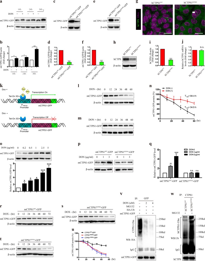
Fig. 1. Forming cytoophidia prolongs the half-life of CTP synthase.
a HEK-293T cells stably expressing mCTPS1-GFP were treated with 4 μg/ml DON for the indicated time, and cell lysates were prepared and analyzed by immunoblotting with anti-GFP antibody. b Quantitative data of mCTPS1-GFP protein level in a. c, e SW480 cells stably expressing mCTPS1WT-GFP, mCTPS1H355A-GFP (c) or mCTPS1R294D-GFP (e) were cultured for 3 days, and then subjected to western blotting analysis with anti-GFP antibody. d, f Quantitative data of mCTPS1-GFP protein level in c and e, respectively. g–i SW480 cells expressing endogenous wild-type CTPS1 (CTPS1WT) and those expressing R294D mutant CTPS1 (CTPS1R294D) were cultured for 3 days, followed by immunostaining (g) or western blotting against CTPS1 (h). i Quantitative data of CTPS1 protein levels in h. j The CTPS1 mRNA levels of SW480 CTPS1WT and SW480 CTPS1R294D+/− cells were analyzed by quantitative reverse transcription PCR (qRT-PCR). k Schematic diagram of TRE3G Tet-On system. l, m HEK-293T cells stably expressing Tet-On mCTPS1-GFP were cultured in the medium containing doxycycline (200 ng/ml) for 24 h, followed by culturing with the doxycycline-free medium without (l) or with (m) DON (4 μg/ml) for the indicated time. Lysates were prepared and analyzed by immunoblotting with appropriate antibodies. n Quantitative data of mCTPS1-GFP protein levels in l and m. o HEK-293T cells stably expressing Tet-On mCTPS1-GFP were cultured in the medium containing doxycycline (200 ng/ml) for 24 h, followed by culturing with the doxycycline-free medium with the indicated concentration of DON for 48 h. Lysates were prepared and analyzed by immunoblotting with appropriate antibodies. p HEK-293T cells stably expressing Tet-On mCTPS1WT-GFP (left three lanes) or Tet-On mCTPS1H355A-GFP (right three lanes) were treated with doxycycline (200 ng/ml) for 24 h, followed by culturing with doxycycline-free medium with the indicated concentration of DON for 48 h. Anti-GFP antibody was used to detect mCTPS1-GFP levels. q Quantitative data of mCTPS1-GFP protein levels in p. r–t SW480 cells stably expressing Tet-On mCTPS1WT-GFP (r), mCTPS1R294D-GFP (s) or mCTPS1H355A-GFP (t) were grown in the medium containing doxycycline (200 ng/ml) for 24 h, followed by culturing with the doxycycline-free medium for the indicated time. Lysates were prepared and subjected to western blotting analysis with anti-GFP antibody. u Quantitative data of mCTPS1-GFP protein levels in r–t. v HEK-293T cells stably expressing mCTPS1-GFP were transfected with HA-Ubiquitin, and then treated with the indicated concentration of DON for 36 h. MG132 (20 μM) was added during the last 16 h of treatment. Lysates prepared were subjected to immunoprecipitation by anti-GFP antibody. Immunoprecipitates were analyzed by immunoblotting using anti-HA antibody. w Wild-type and CTPS1 R294D mutant SW480 cells were treated with MG132 for 16 h. Lysates were prepared and subjected to immunoprecipitation by anti-CTPS1 antibody. Immunoprecipitates were analyzed by immunoblotting using anti-ubiquitin antibody. Mean ± S.E.M, *P < 0.05; **P < 0.01; ***P < 0.001; ****P < 0.0001 versus control. Scale bars = 20 μm. One of three to five similar experiments is shown. DAPI, 40,6-diamidino-2-phenylindole; DON, 6-diazo-5-oxo-l-norleucine

