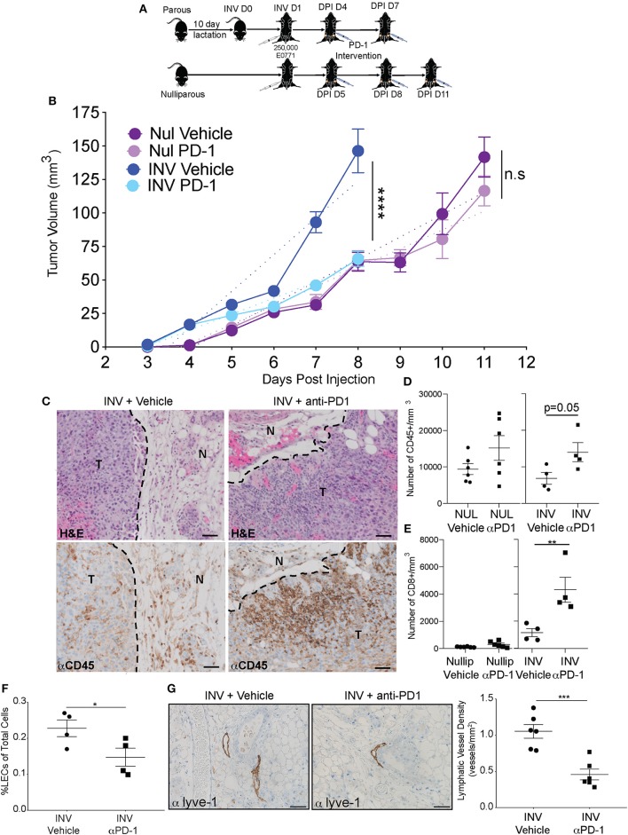
Figure 4.
Anti-PD-1 treatment reduces tumor growth, enhances immune infiltration, and reduces lymphatic vessels in a model of post-partum breast cancer (A) C57/Bl6 mice were bred and allowed to lactate for 10 days before pups were removed to initiate mammary gland involution (parous/involution, n = 6); age-matched nulliparous controls were used (n = 6). Parous animals were injected with 250,000 E0771 tumor cells at Inv D1 (involution group) or into nulliparous animals (nulliparous group). Once tumors became measurable (involution = 4 days post injection (DPI); nulliparous=DPI D5), anti-PD-1 intervention was administered and continued every third day. E0771 mammary tumor growth curves from nulliparous and involution group C57Bl/6 mice treated with vehicle or anti-PD-1 are shown in (B). Results are representative from two independent studies. Dotted lines represent the slope of the tumor growth. (C) Representative images of H&E analysis and immunohistochemistry for CD45 (brown) in fixed tumor tissue to identify tumor infiltrating lymphocytes after anti-PD-1 treatment compared to vehicle controls, T = tumor and N = normal. CD45+ area was 9.99% of tumor area for involution with vehicle and 22.62% of tumor area for involution with anti-PD-1 treatment. Scale bars are 50 microns. Number of (D) CD45+ and (E) CD8+ cells per area in tumors from B quantified by flow cytometric analysis. (F) % LECs of total in tumors from B quantified by flow cytometry. (G) Representative images of Lyve-1 stained fixed tumor adjacent tissues and quantitation of Lyve-1+ vessels per area in involution group tumors +/- PD-1 treatment. Scale bars are 100 microns. *p < 0.05; **p < 0.01, ***p < 0.001, ****p < 0.0001.

