Figure 2.
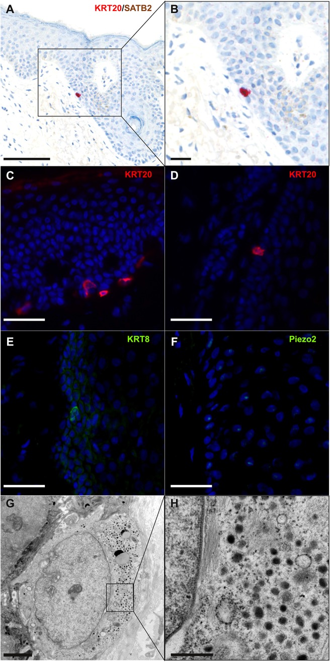
Immunohistochemical and ultrastructural features of physiological Merkel cells: immunohistochemical staining of normal skin (A,B) revealed one Merkel cell located in the infundibulum of a hair follicle and coexpressing cytokeratin 20 (cytoplasmic expression in red) and SATB2 (nuclear expression in brown) (bar = 100 and 50 μm for A,B). Immunofluorescence staining of healthy skin revealed some Merkel cells expressing cytokeratin 20 (C,D), cytokeratin 8 (E) and Piezo2 (F) in the epidermis (C) and in hair follicles (D–F) (bar = 40 μm for C–F). Electron microscopy of a Merkel cell (G,H) revealed numerous dense-core granules (bars = 2 and 0.5 μm for G,H, respectively). A cropped region is shown in the inset (H).

