
Figure 2.
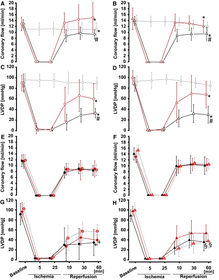
Coronary flow and LVDP in isolated perfused rat hearts. Values are means ± SD. Circles represent female, and triangles represent male rat hearts. (A, B) Coronary flow and (C, D) LVDP in hearts with: local ischemic preconditioning (red: IPC+GI/R), time‐matched perfusion (black: TP+GI/R), time control (grey: time control), (E, F) coronary flow and (G, H) LVDP in hearts with: placebo protocol (black: placebo+GI/R), 1‐RIPC (grey with red margin: 1‐RIPC+GI/R), 2‐RIPC (red: 2‐RIPC+GI/R); *P < 0.05 versus time‐control, respectively, #P < 0.05 versus IPC+GI/R, respectively +P < 0.05 versus 1‐RIPC, §P < 0.05 versus 2‐RIPC (two‐way ANOVA).