Figure 1.
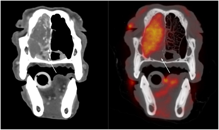
Transverse post-contrast CT (left) and fused 18F-FDG PET/CT (right) images through the caudal aspect of the nasal cavity of a 10-year-old male castrated standard poodle presented for staging of nasal adenocarcinoma. There is a large soft tissue mass filling the left side of the nasal cavity and resulting in destruction of the nasal turbinates and invasion of the maxillary recess. The soft tissue opacity extends ventrally into the rostral aspect of the nasopharynx. The opacity in the nasopharynx is strongly contrast enhancing on the CT image but does not show FDG uptake whereas the rest of the mass demonstrates strong FDG uptake. This suggests that the tissue in the nasopharynx differs from the bulk of the tumor and might represent edematous nasopharyngeal mucosa rather than neoplastic tissue.

