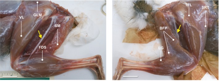
Figure 1.

Muscle topography of Sylvilagus floridanus. Lateral view photographs of adult R41 (left) and adult R39 (right) hind limbs during dissection. The superficial m. biceps femoris vertebral head is removed to expose the sciatic nerve (yellow arrows) and deep musculature in both photos. The m. biceps femoris pelvic head is also removed (left) to uncover the ankle extensors and digital flexors. Pins are placed in the approximate center of rotation of the hip and knee joints for measurement of muscle moment arms (r m). Selected muscles are labeled in each photo for absolute orientation. BFP, m. biceps femoris pelvic head; FDS, m. flexor digitorum superficialis; GLM, m. gluteus medialis; LG, m. gastrocnemius lateral head; SM, m. semimembranosus; SRT, m. sartorius; TFL, m. tensor fascia latae; and VL, m. vastus lateralis. Scale bar: 1.5 cm.
