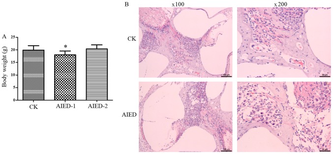
Figure 1.
Construction of autoimmune inner ear disease model mice. (A) Measurement of body weight in each group. (B) Histopathological analysis of the cochlea via standard hematoxylin and eosin staining. 2, for two weeks. *P<0.05 vs. CK group. CK, control; AIED, autoimmune inner ear disease; AIED-1, 1 week AIED model; AIED-2, 2 week AIED model.

