Figure 2.
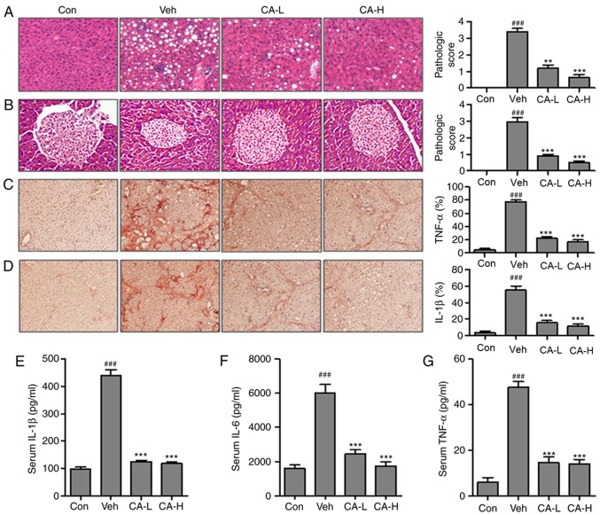
CA decreases the inflammatory response in HFD mice. Hematoxylin and eosin staining of (A) liver and (B) pancreas indicated the pathological score in different experimental groups (magnification, ×50). Immunohistochemical analysis of (C) TNF-α and (D) IL-1β in the liver of HFD mice and percentage of positive cells. Serum levels of (E) IL-1β, (F) IL-6 and (G) TNF-α. Data are expressed as the means ± SEM. n=10 in each group. ###P<0.001 vs. Con; **P<0.01 and ***P<0.001 vs. Veh. CA, carnosic acid; CA-H, HFD mice treated with 20 mg/kg CA; CA-L, HFD mice treated with 10 mg/kg CA; Con, Control group; HFD, high-fat diet; IL, interleukin; TNF-α, tumor necrosis factor-α; Veh, HFD mice.

