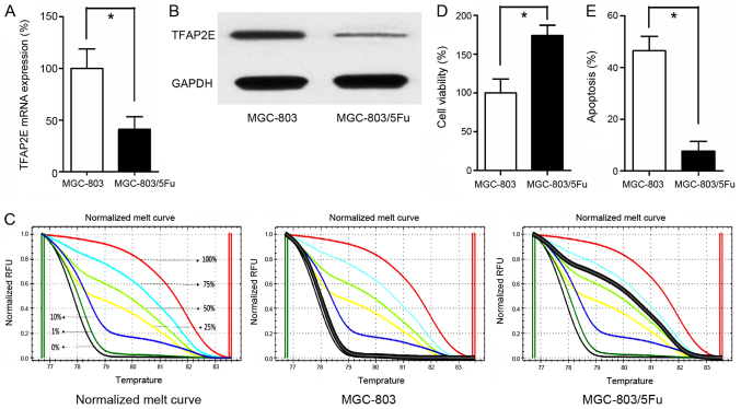
Figure 1.
TFAP2E methylation status and expression level in MGC-803 and MGC-803/5-FU cells. (A) qRT-PCR detection of TFAP2E mRNA expression in MGC-803 and MGC-803/5-FU cells, GAPDH was assayed as an internal control. (B) Representative western blotting results. (C) MS-HRM analysis for TFAP2E methylation, including standard curves. (D and E) Cell viability and apoptosis were assessed by cell cytotoxicity and apoptosis assays, respectively. All the results were the average from at least 3 independent experiments. Mean ± SD; *P<0.05, two-tailed Student's t-test. TFAP2E, transcription factor activating enhancer-binding protein 2e; 5-FU, 5-fluorouracil; MS-HRM, methylation-sensitive high-resolution melting; SD, standard deviation.

