Figure 2.
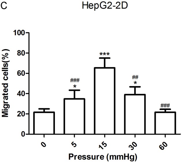
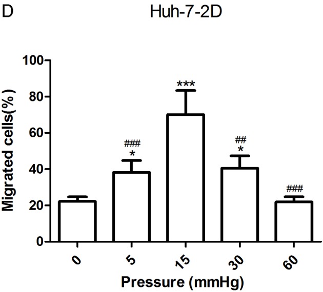
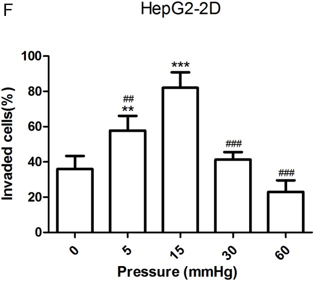
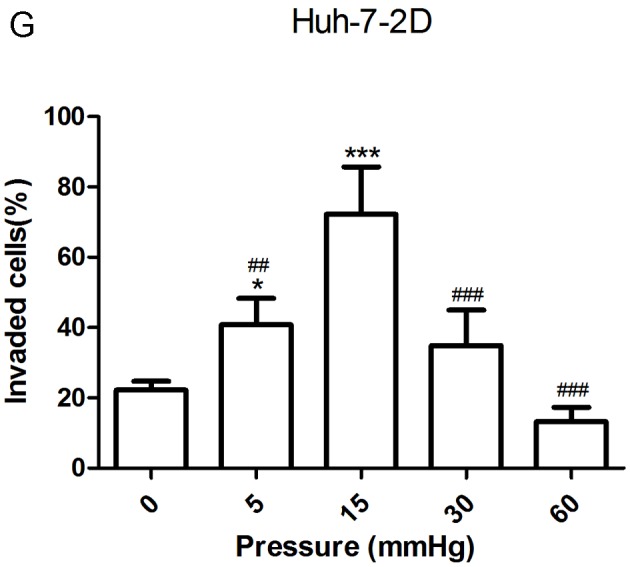
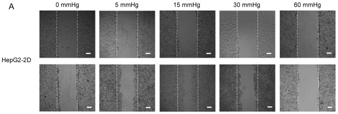
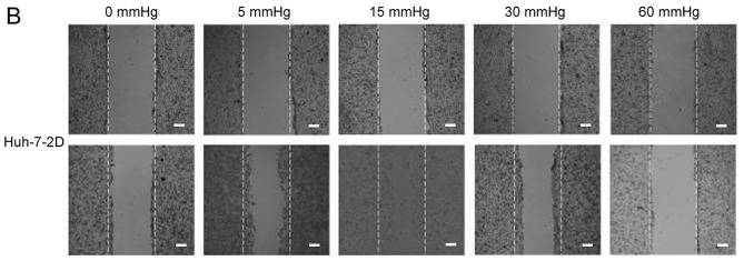
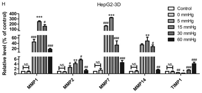
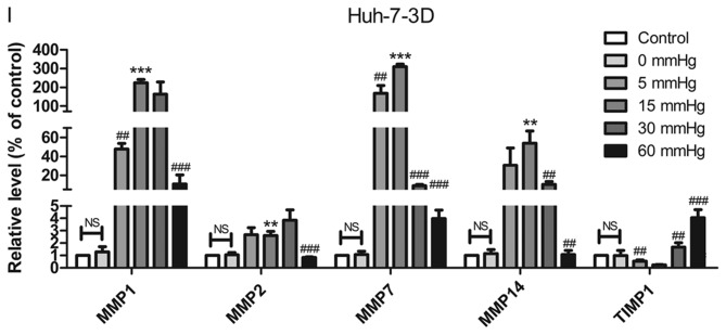
Effect of pressure on HepG2 and Huh-7 liver cancer cell migration and invasion in vitro. (A) HepG2 and (B) Huh-7 confluent cell monolayers were wounded and cultured under different levels of pressure (0, 5, 15, 30, 60 mmHg) for 24 h (magnification, ×100); (C) HepG2 and (D) Huh-7 percentage of migration was calculated. (E) Representative images of Transwell assay results showing cells stained with 0.1% crystal violet solution (magnification, ×100). Effect of pressure on HepG2 and Huh-7 liver cancer cell migration and invasion in vitro. The assay lasted 24 h, and after fixing and staining, the percentage of (F) HepG2 and (G) Huh-7 cells on the lower chamber was calculated; 10% acetic acid was used to elute the stained inserts to detect the percentage of invaded cells. mRNA expression levels of MMP1, MMP2, MMP7, MMP14 and TIMP1 in 3D-cultured (H) HepG2 and (I) Huh-7 cells exposed to different levels of pressure (0, 5, 15, 30, 60 mmHg) for 24 h. All data are expressed as the means ± standard deviation. The data were analyzed using ANOVA, followed by the least significant difference post hoc test. *P<0.05, **P<0.01 and ***P<0.001 vs. 0 mmHg. #P<0.05, ##P<0.01 and ###P<0.001 vs. 15 mmHg. 2D, 2-dimensional; 3D, 3-dimensional; MMP, matrix metalloproteinase; TIMP, tissue inhibitor of metalloproteinases; NS, not significant.

