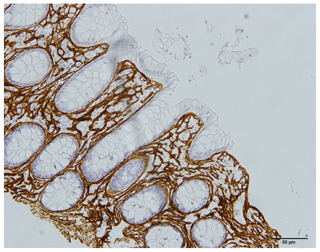
Figure 5.

Photomicrograph taken from the colon of a 42-year-old male who fulfilled the Rome III criteria. A histopathological examination revealed the presence of collagenous colitis. The section showed positive immunostaining for collagen III. Scale bar, 50 µm.
