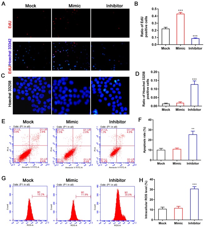
Figure 2.
Effect of miR-144 on BMSC proliferation and apoptosis. (A) EdU staining was used to evaluate and the effect of miR-144 on BMSC proliferation (magnification, ×100). (B) Ratio of Edu-positive cells to total cells. (C) Effect of miR-144 on the nuclear morphological features of BMSCs was observed following Hoechst 33258 staining (magnification, ×200). (D) Ratio of Hoechst 33258-positive cells to total cells. (E) Effect of miR-144 on BMSC apoptosis was detected by flow cytometry and (F) quantified. (G) Intracellular ROS in BMSCs were detected by flow cytometry and (H) quantified. The group of BMSCs transfected with miR-144 mimic or the miR-144 inhibitor were compared with BMSCs in the mock groups. Data represent the mean ± SEM. **P<0.01 and ***P<0.001. miR, microRNA; BMSCs, bone marrow-derived mesenchymal stem cells; ROS, reactive oxygen species.

