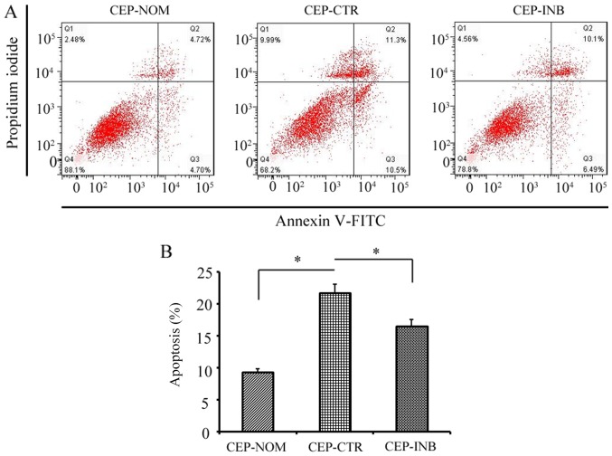
Figure 3.
Evaluation of the apoptosis of CEP cells following LiCl treatment. (A) Representative graphs of flow cytometric analysis. (B) Apoptotic rate of CEP cells was significantly higher following LiCl treatment, whereas inhibiting Wnt/β-catenin signalling protected against cellular apoptosis. The results are presented as the percentage of cell apoptosis. *P<0.05 CEP-CTR vs. CEP-NOM and CEP-INB. CEP-NOM, cells cultured under normal conditions; CEP-CTR, cells treated with LiCl; CEP-INB, cells treated with LiCl and a β-catenin inhibitor; CEP, cartilage endplate; LiCl, lithium chloride; FITC, fluorescein isothiocyanate.

