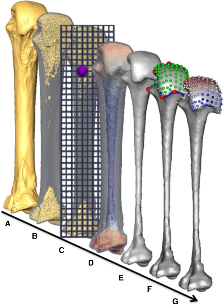
Figure 2.

Methodological stages of metacarpal trabecular analysis, shown in a third metacarpal as an example: (A) isosurface model, (B) segmented trabecular structure inside cortical shell, (C) diagram of the background grid and one of the VOIs at a vertex (purple), (D) volume mesh coloured by BV/TV (0–45%), (E) smoothed trabecular surface mesh, (F) surface landmarks (anatomical = red, semi‐sliding landmarks on curves = blue and on surfaces = green), (G) RBV/TV interpolated to each surface landmark.
