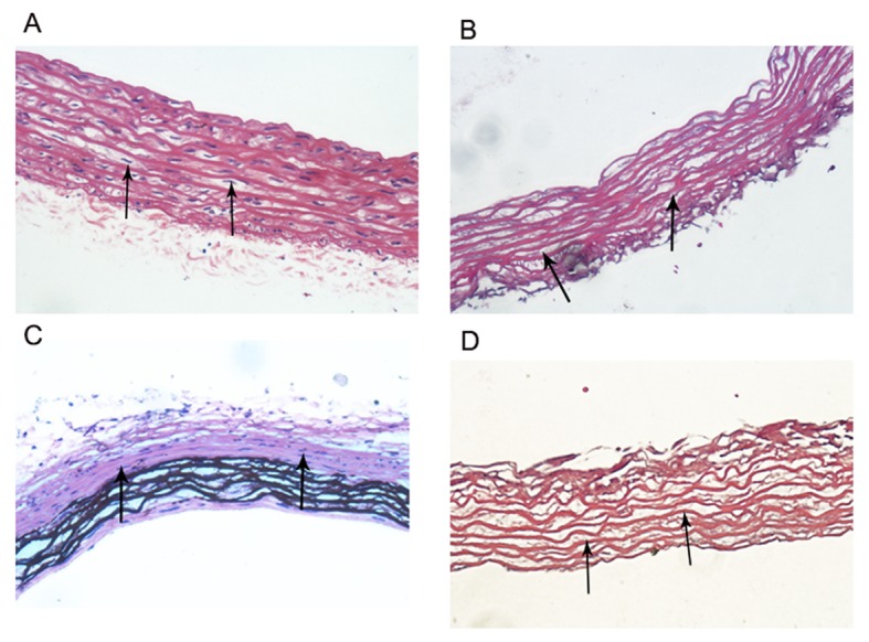
Figure 2.

Biological scaffolds. (A) There were numerous nuclei in normal arteries (black arrows; magnification, ×40). (B) There were no nuclei in the normal arterial decellularized scaffolds; however, the elastic fiber structure was complete and orderly arranged (black arrows; magnification, ×40). (C) There were numerous nuclei in calcified arteries (white arrows; magnification, ×40). (D) There were no nuclei in the calcified arterial decellularized scaffolds, however, the elastic fiber structure was complete and orderly arranged (black arrows; magnification, ×40).
