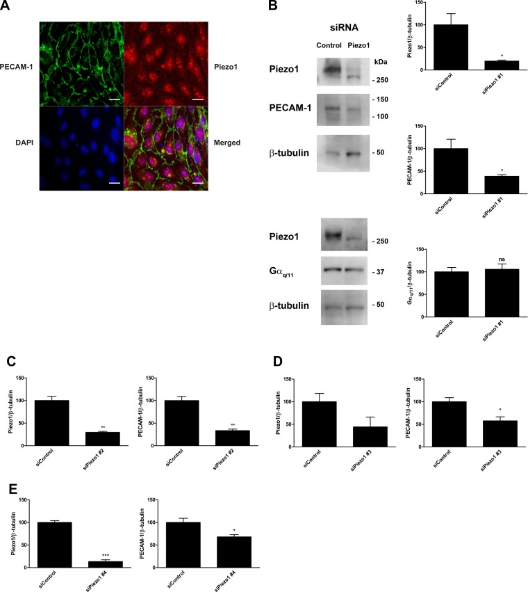
Fig. 3.
Piezo1 protein localization and the effects of different Piezo1 siRNA oligonucleotides on Piezo1 and PECAM-1 expression in endothelial cells. A: human coronary artery endothelial cells (HCAECs) were fixed, blocked, and immunostained with an anti-Piezo1 primary antibody followed by an Alexa Fluor 568-conjugated secondary antibody (red). Cells were counterstained with a fluorescein-conjugated anti-PECAM-1 antibody (green) and mounted with medium containing DAPI (blue). A representative image from 3 independent experiments is shown. Scale bar, 20 μm. B: HCAECs were transfected with either control siRNA or siRNA against Piezo1 and lysed after 48 h. Immunoblotting was performed on cell lysates using antibodies against Piezo1, PECAM-1, Gαq/11, and β-tubulin. Representative blots from 3 independent experiments are shown. Bar graphs represent the ratios of Piezo1, PECAM-1, and Gαq/11 expression to β-tubulin expression with control siRNA set to 100%. Error bars indicate SE. *P < 0.05. C–E: HCAECs were transfected with either control siRNA or three different siRNAs against Piezo1: siPiezo1 #2 (C), siPiezo1 #3 (D), and Piezo1 #4 (E). At 48 h after transfection, cells were harvested and lysed. Immunoblotting was performed on cell lysates using antibodies against Piezo1, PECAM-1, and β-tubulin. Bar graphs reflect the quantification of at least 3 independent experiments and are presented as the ratios of Piezo1 and PECAM-1 expression to β-tubulin expression with control siRNA set to 100%. Error bars indicate SE. *P < 0.05; **P < 0.01; ***P < 0.001.

