Fig. 7.
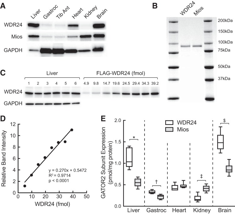
Western blot analysis of WDR24 and Mios protein expression in liver, gastrocnemius, tibialis anterior, heart, kidney, and brain of freely fed rats. A: representative Western blot analysis of WDR24, Mios, and GAPDH expression in the same samples analyzed in Fig. 3. B: FLAG-tagged WDR24 and Mios were individually expressed in and purified from HEK293T cells as described under materials and methods. Purified proteins (2 µg) were resolved by SDS-PAGE, and the gel was stained with SimplyBlue SafeStain. Ratio, band intensity relative to WDR24. C: Western blot analysis of WDR24 and GAPDH expression in liver (n = 6) and various amounts of purified FLAG-WDR24. For GAPDH, only the liver samples were analyzed. D: representative standard curve for FLAG-WDR24. E: quantification of Western blot analysis of various tissues. Values represent means ± SE (n = 6). Method used to quantify WDR24 shown in C and D was also used to quantify Mios using the respective purified protein (B). *WDR24 and Mios expression in liver differ significantly, P < 0.005; †WDR24 and Mios expression in gastrocnemius differ significantly, P < 0.005; ‡WDR24 and Mios expression in kidney differ significantly, P < 0.005 kidney; and WDR24 and Mios expression in brain differ significantly, $P < 0.005. Gastroc, gastrocnemius; GATOR2, GAP activity toward Rags 2; Mios, meiosis regulator for oocyte development; Tib Ant, tibialis anterior; WDR24, WD repeat-domain-containing protein 24.

