Fig. 3.
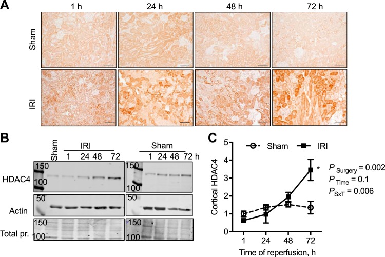
Time course in changes in histone deacetylase-4 (HDAC4) expression and abundance in the male mouse kidney cortex after 30 min of bilateral ischemia followed by 1–72 h of reperfusion (IRI) or sham surgery. A: HDAC4 was immunolocalized to the proximal tubule in the kidney cortex. Scale bar = 40 μm. B: representative Western blots of cortical lysates and HDAC4 abundance with β-actin and total protein (pr.; Coomassie blue-stained membranes) to demonstrate equal loading. C: relative quantification of HDAC4 cortical abundance normalized to 1-h sham samples. n = 3–4 Animals per time point. Two-factor ANOVA, *P < 0.05 compared with sham from Šídák multiple-comparison post hoc analysis. 50, 100, and 150, 50-, 100-, and 150-kDa molecular mass markers; PSxT, PSurgery×Time.

