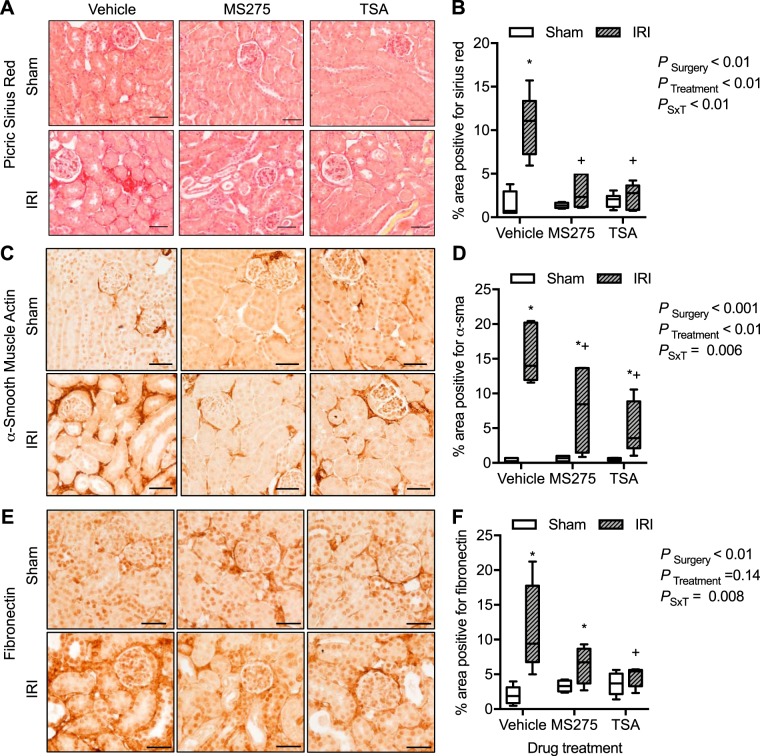
Fig. 8.
Kidney cortical interstitial collagen and extracellular matrix proteins from mice implanted with intraperitoneal osmotic minipumps to deliver continuously vehicle (acetic acid, pH > 5), MS275, the class I histone deacetylase (HDAC) inhibitor (20 mg·kg−1·day−1), or trichostatin A (TSA; 1 mg·kg−1·day−1), the pan-HDAC inhibitor, 3 days before sham surgery or 27 min of bilateral ischemia (ischemia-reperfusion injury, IRI). Samples were obtained at 72 h of reperfusion. Shown are Sirius red staining in the interstitium (A) and percentage of area positive for Sirius red (B). Also shown are expression of α-smooth muscle actin (α-SMA; C) and percentage of area positive for α-SMA (D). Expression of fibronectin (E) and percentage of area positive for fibronectin (F) are shown as well. Box plots ± minimum and maximum are reported. n = 4–7 Animals; see materials and methods for exact sample size per group. Two-factor ANOVA, *P < 0.05 compared with sham, +P < 0.05 compared with IRI vehicle from Tukey post hoc analysis. G, glomerulus; PSxT, PSurgery×Treatment. Scale bar = 40 μm.

