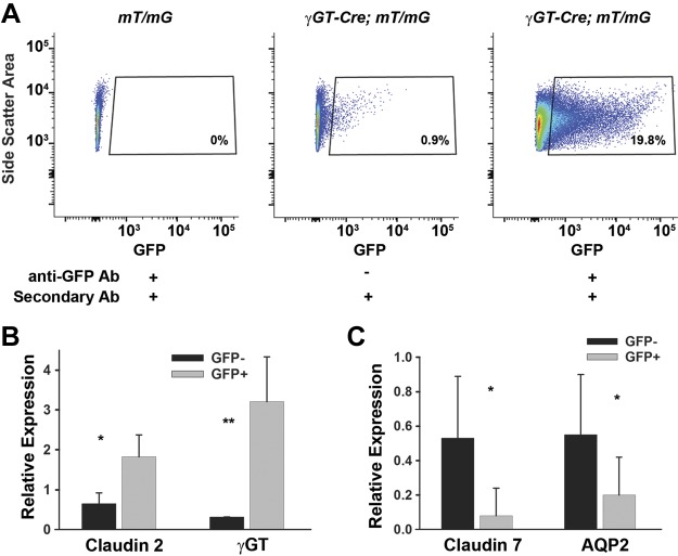
Fig. 3.
Antibody enhances green fluorescent protein (GFP) detection and validation of the GFP+ population. A: mice lacking γ-glutamyltransferase 1 (γGT)-Cre were used as negative controls. γGT-Cre;mT/mG mice without the GFP antibody had some positive cells, but this was greatly enhanced with the use of a GFP antibody. B: to validate that GFP+ cells are proximal tubule cells, we sorted out the GFP+ population, isolated mRNA, and performed quantitative PCR for proximal tubule-specific genes (claudin 2 and γGT) and for distal tubule genes [claudin 7 and aquaporin 2 (AQP2)]. Gapdh was used as a loading control. The value of GFP− was normalized to 1 in 1 mouse with other values relative to this mouse. Error bars represent SD for a total of 4 mice. *P < 0.05 and **P < 0.01, statistically significant differences in gene expression. The gating on GFP+ cell population in A is from 1 representative mouse.

