Fig. 4.
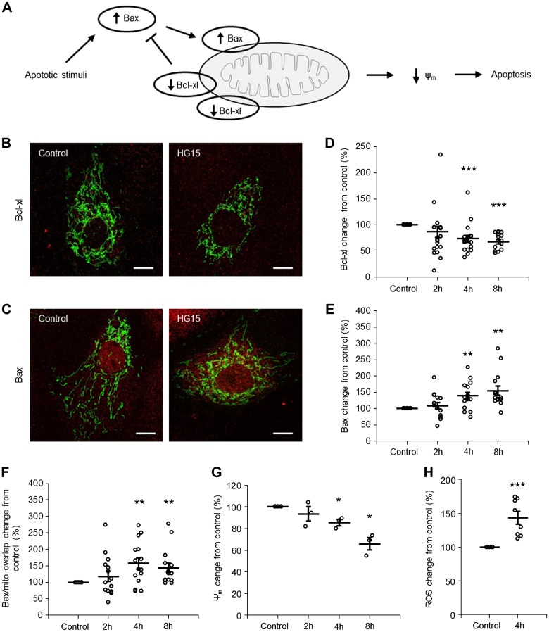
High glucose triggers apoptosis via the mitochondrial pathway in a time-dependent manner in proximal tubule cell (PTCs). A: cartoon illustrating activation of the mitochondrial apoptotic pathway. Under normal conditions, there is a balance between Bcl-xL and Bax, preventing apoptosis. When an apoptotic stimulus, i.e., high glucose, activates the intrinsic apoptotic pathway, the balance between Bax and Bcl-xL is disrupted, which leads to mitochondrial dysfunction [decreased mitochondrial membrane potential (Δψm)] and apoptosis. B and C: immunofluorescence staining for Bcl-xL (B) and Bax (C) expression (red) in PTCs incubated with control (5.6 mM) or 15 mM glucose (HG15)-containing medium for 8 h. Mitochondria are shown in green. Scale bars = 10 µm. D−F: quantification of Bcl-xL abundance (D), Bax abundance (E), and Bax accumulation on mitochondria (F) in PTCs incubated with control or 15 mM glucose-containing medium for 2, 4, and 8 h. Data are expressed as means ± SE; n = 15 coverslips from 5 individual cell preparations. G: quantification of Δψm in PTCs incubated with control or 15 mM glucose-containing medium for 2, 4, and 8 h. Data are expressed as means ± SE; n = 3 coverslips from 3 individual cell preparations. H: quantification of ROS in PTCs incubated with control or 15 mM glucose-containing medium for 4 h. Data are expressed as means ± SE; n = 8 coverslips from 2 individual cell preparations. *P < 0.05; **P < 0.01; ***P < 0.001.

