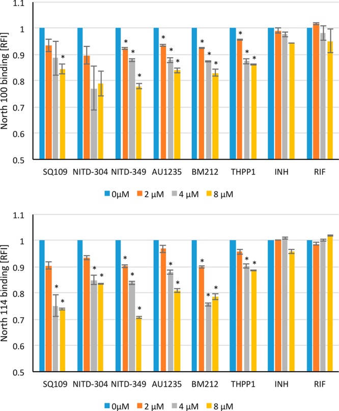
Figure 4.

Flow-cytometry-based competition binding assay using intact Msmg cells expressing MmpL3tb. Flow-cytometry-based competition binding assay was performed in an Msmg mmpL3 deletion mutant expressing the wild-type mmpL3tb gene fused to gfp (MsmgΔmmpL3/pMVGH1-mmpL3tb). Cells were labeled with 4 μM North 100 (top graph) or North 114 (bottom graph) and subsequently treated with increasing concentrations of the inhibitors as described in the Methods. The concentrations of inhibitors are indicated under the x-axis. Shown on the y-axis are the MFI of the bacilli from each treatment group expressed relative to that of bacilli not treated with any inhibitor (relative fluorescence value [RFI] arbitrarily set to 1). MFIs were determined by analyzing 10 000 bacilli under each condition. The data reported are mean values ± SD of technical duplicates and are representative of at least three independent experiments. Asterisks denote statistically significant decreases in fluorescence intensity between no inhibitor controls (blue bars) and bacilli cotreated with North 100 or North 114 and various concentrations of the inhibitors pursuant to the Student’s t-test (P < 0.05).
