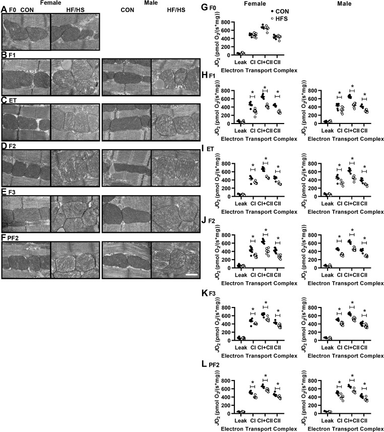
Fig. 2.
Altered mitochondrial morphology and function in descendants of HFS-fed dams. A–F: representative transmission electron microscopy images at ×25,000 magnification of myocardial sections from mice of the indicated generations as in Fig. 1; n = 3/group. Scale bar = 500 nm. G–L: high-resolution respirometry was performed to measure oxygen consumption [volume-specific oxygen flux (JO2)] in isolated, permeabilized cardiac fiber bundles in the presence of malate/pyruvate/glutamate (Leak), ADP (complex I), succinate (complex I+II), and rotenone (complex II); n = 8/group. All data are expressed as means ± SE. *P < 0.05 by Student’s t-test between Con and HFS groups, within sex and generation.

