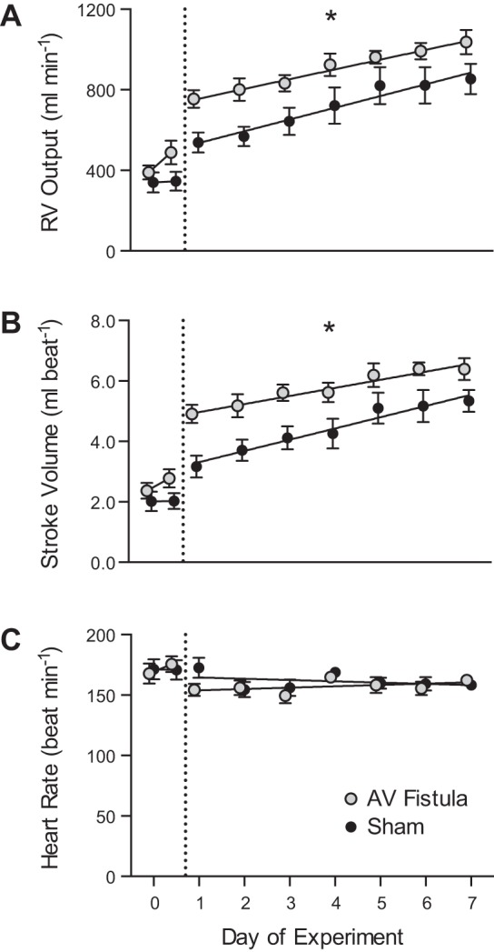
Fig. 1.

Hemodynamics in experimental fetal right carotid artery-jugular vein (AV) fistula. A–C: right ventricular (RV) output, stroke volume, and heart rate in fetuses with AV fistula and sham controls. Points to the left of the dashed vertical line represent data collected during surgery (day 0) in the anesthetized state. For the AV fistula group, the day 0 point on the left is with the fistula closed, and the point on the right is with the fistula open. For the sham group, the day 0 point on the left is with the fistula closed, and the point on the right is with the fistula ligated. Data for days 1–7 were obtained from unanesthetized fetuses. Values are means ± SE. All RV output and RV stroke volume values on days 1–7 were higher for the AV fistula than the sham group: *P < 0.0001. Comparative slopes between AV fistula and sham fetuses for the relationship for RV output, stroke volume, and heart rate study as a function of time were not different: P = 0.53, P = 0.28, and P = 0.16, respectively.
