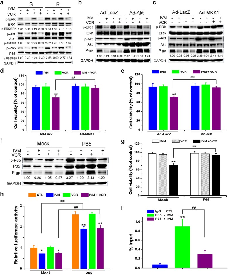
Fig. 7.
Ivermectin decreased P-gp expression through inhibiting ERK/Akt and NF-κB activation. a The expression levels of the proteins of the VCR-resistant/sensitive HCT-8 cells treated with 25 nM vincristine (VCR) and/or 3 μM ivermectin (IVM) for 48 h were determined. b-h Expression levels of the proteins (b, c, f), the cell viability (d, e, g), and the relative MDR1 promoter activity (h) of the VCR-resistant HCT-8 cells infected by recombinant adenovirus expressing HA-tagged constitutively active Akt (Ad-Akt-myr) (b and e) or by the flag-tagged constitutively active MKK1 (Ad-MKK1-R4F) (c and d), or transfected with plasmid pcDNA3.1(+)-P65, treated with 25 nM VCR and/or 3 μM IVM for 48 h were determined. i Chromatin IP was carried out with IgG (negative control) and anti-P65 antibody. Q-PCR result for MDR1 promoter region was shown as the percentage of input DNA. Cell viability was detected by MTT assay and the protein expression levels were detected by Western blotting analysis using GAPDH as internal control. Relative MDR1 promoter activity was determined by Gaussia luciferase activity normalized to the transfection control, i.e., secreted alkaline phosphatase (SeAP). Cells treated with recombinant adenovirus expressing Ad-LacZ or with empty vector pcDNA3.1(+) (mock) serve as control. Abbreviations: CTL, control; IVM, ivermectin; VCR, vincristine; S, vincristine-sensitive cells; R, vincristine-resistant cells. Western blots in a-c and f are representative of two independent experiments. Data in d, e, and g represent the percentage of respective control values (mean ± SD, n = 5). Data in h are expressed as fold change of the activity over the control from the mock group (mean ± SD, n = 3). Data in i are expressed as the mean ± SD (n = 4). Statistical significances in d, e, and g-i were determined using one-way ANOVA followed by Dunnett’s test. *P < 0.05, **P < 0.01, compared with the respective controls; ##P < 0.01, comparison between the two columns

