Fig. 2.
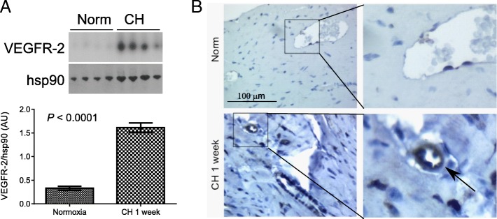
Right ventricular VEGFR-2 expression in CH-PH. VEGFR-2 expression is increased in the RV of mice exposed to CH-PH for 1 week, as demonstrated by western blot (a). Graphs denote relative expression (normalized to RV hsp90 expression) of n = 4 animals/group. Immunohistochemistry (b) identifies perivascular staining in RV sections from mice exposed to CH-PH, consistent with an endothelial cell source for observed increases in tissue VEGFR-2. Error bars represent SEM. P-value is for Student’s t-test

