Fig. 4.

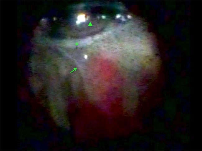
Endoscopic view in a patient with chronic retinal detachment and hypotony showing a tractional retinal detachment (arrow) connected to an epiciliary membrane (asterisk). An air bubble is located superiorly (arrowhead)

Endoscopic view in a patient with chronic retinal detachment and hypotony showing a tractional retinal detachment (arrow) connected to an epiciliary membrane (asterisk). An air bubble is located superiorly (arrowhead)