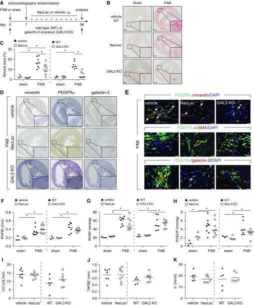
Figure 2.
Pharmacologic inhibition and genetic deletion of galectin-3 ameliorates the progression of right ventricle (RV) fibrosis. (A) Schematic representation of the experimental setup. Hearts were collected from inhibitor (NacLac), knockout (galectin-3 homozygous knockout mice), or vehicle/wild-type treated mice 21 days after randomization and treatment. (B) Representative Sirius red staining. Scale bars, 500 μm. (C) Quantification of RV fibrosis. (D) Representative immunohistochemical staining of the murine hearts against vimentin, PDGFRα (platelet-derived growth factor receptor-α), and galectin-3 (n = 3). Scale bars, 500 μm. (E) Representative immunofluorescence staining of the RV fibrotic regions against vimentin, PDGFRα, αSMA (α-smooth muscle actin), and galectin-3 (n = 2). Arrows depict cells showing colocalization of PDGFRα and vimentin or galectin-3. Scale bars, 20 μm. (F) Echocardiographic assessment of RV free wall thickness. Invasive hemodynamic measurement of (G) RV systolic pressure and (H) RV end diastolic pressure. Echocardiographic assessment of (I) cardiac output, (J) tricuspid annular plane systolic excursion, and (K) early peak of RV relaxation velocity. Two-way ANOVA with Bonferroni post hoc test, *P < 0.05. CO = cardiac output; e′ = RV relaxation velocity; GAL3 KO = galectin-3 homozygous knockout mice; i.p. = intraperitoneal; NacLac = N-acetyllactosamine; PAB = pulmonary artery banding; RVEDP = RV end diastolic pressure; RVFW = RV free wall thickness; RVSP = RV systolic pressure; TAPSE =tricuspid annular plane systolic excursion; WT = wild type.

