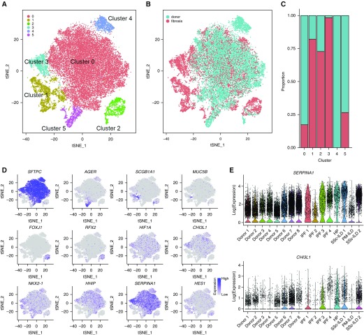
Figure 6.
Distinct populations of alveolar epithelial cells emerge during fibrosis. (A) Six clusters were identified after epithelial cells from each of eight normal and eight fibrotic lungs were combined and clustered. (B and C) Relative contributions of epithelial cells from normal and fibrotic lungs to each cluster as shown by t-distributed Stochastic Neighbor Embedding plot and by bar plots. (D) Feature plots demonstrating differential expression of selected epithelial marker genes: SFTPC (alveolar type II cells), AGER (alveolar type I cells), SCGB1A1 (club cells), FOXJ1, and RFX2 (ciliated airway epithelial cells). Also shown are genes implicated in pulmonary fibrosis (HIF1A, CHI3L1, NKX2-1, HHIP, FASN, and HES1). (E) Violin plots representing heterogeneity in expression of SERPINA1 and CHI3L1 in epithelial cells from normal and fibrotic lungs. For definition of abbreviations, see Figure 5.

