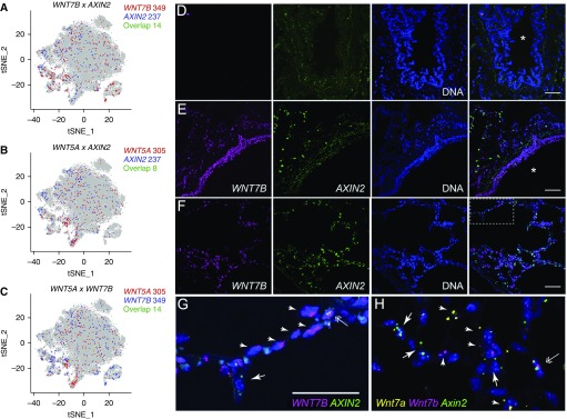
Figure 8.
Wnt secretion and response are restricted to distinct nonoverlapping epithelial cells. (A–C) Expression of WNT7B, WNT5A, and AXIN2 in epithelial cells from normal and fibrotic lungs by single-cell RNA-Seq. Total number of single-positive or double-positive cells is indicated. (D and E) Fluorescence RNA in situ hybridization with amplification reveals Wnt-expresser and Wnt-responder cells in the proximal airways of human lung. Images were obtained from human donor lung sections incubated without (D) or with designated target probes, WNT7B (magenta) and AXIN2 (green) (E–G). Nuclei are stained with Hoechst (blue) and overlay images are shown on the far right. Note WNT7B is abundantly detected in small airway epithelial cells (E), consistent with its prominent detection in club cells by single-cell RNA-Seq. AXIN2 is not similarly enriched in club cells and is only specifically detected in alveolar regions (F) under higher magnification (G is a higher-magnification image of the boxed region in F). (H) High magnification of mouse lung alveoli subjected to fluorescence RNA in situ hybridization with amplification with probes for Wnt7a (yellow), Wnt7b (magenta), and Axin2 (green). Arrows indicate cells that are only positive for Axin2; arrowheads indicate specified Wnts. Double arrows indicate cells that may express both Wnts and Axin2, although quantification suggests cells that highly express Wnts and Axin2 are rare (Figure E9D). Asterisks indicate airway lumen. Scale bars, 50 μm. tSNE = t-distributed Stochastic Neighbor Embedding.

