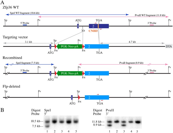
FIG 3.
Scheme of Zfp36 gene targeting in mice. (A) Schematic representation of the mouse Zfp36 locus, the targeting vector, the homologously recombined features in the mutant allele, and after the Flp-mediated deletion of the PGK-Neo positive-selection cassette. The blue or pink arrows indicate the sizes of the SpeI- or PvuII-digested fragments from WT Zfp36 and the correctly recombined mutant locus. The gray arrows indicate the sizes of the left or right homologous recombination arms in the targeting vector. The half-headed red arrows represent the genotyping primers. Sp, SpeI; Pv, PvuII; Xb, XbaI; 1, exon 1; 2, exon 2; CNBD, CNOT1 binding domain; Frt, flippase recognition site; DTA, MC1-DTA negative-selection marker. (B) Sample Southern blots show SpeI- or PvuII-digested fragments of genomic DNA from five ES cell clones, including one (lanes 1) with the correctly recombined Zfp36 mutant locus, showing hybridization with the 5′ and 3′ probes, respectively. Lanes 3 represent a clone with apparent partial integration at the Zfp36 locus.

