FIG 2.
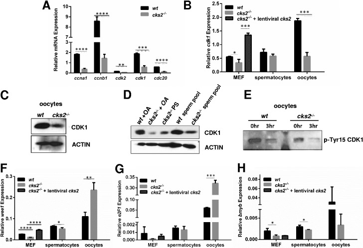
Reduced expression of MPF components cdk1, ccna1, and ccnb1 in cks2−/− oocytes. (A) qRT-PCR analysis showing reduced expression of MPF components in cks2−/− oocytes. Expression of cdk2 and cdc20 is also shown. Data represent the mean ± SD. Significance was determined by Student's t test. ****, P < 0.0001; ***, P < 0.001; **, P < 0.01. (B) Reduced cdk1 expression in cks2−/− oocytes, spermatocytes, and MEFs. Expression of cks2 in cks2−/− MEFs restored the expression of cdk1 to the wt level. Data represent the mean ± SD. Significance was determined by Student's t test. ***, P < 0.001; *, P < 0.05. (C) Immunoblots confirming reduced CDK1 in cks2−/− oocytes. Actin is shown as a loading control. (D) Immunoblots showing reduced CDK1 in cks2−/− spermatocytes. (E) Immunoblot of phosphorylated-Tyr15 (p-Tyr15) CDK1 in wt and cks2−/− oocytes. GV stage oocytes were analyzed at 3 h post-meiotic resumption. Normalized p-Tyr15 CDK1 is 1:1.40 wt/cks2−/−. (F to H) qRT-PCR analysis of wee1 (F), e2f1 (G), and bmyb (H) in wt and cks2−/− oocytes, spermatocytes, and MEFs. wee1 and e2f1 expression was restored in cks2−/− MEFs by enforced cks2 expression. Data represent the mean ± SD. Significance was determined by Student's t test. ****, P < 0.0001; ***, P < 0.001; **, P < 0.01; *, P < 0.05.

