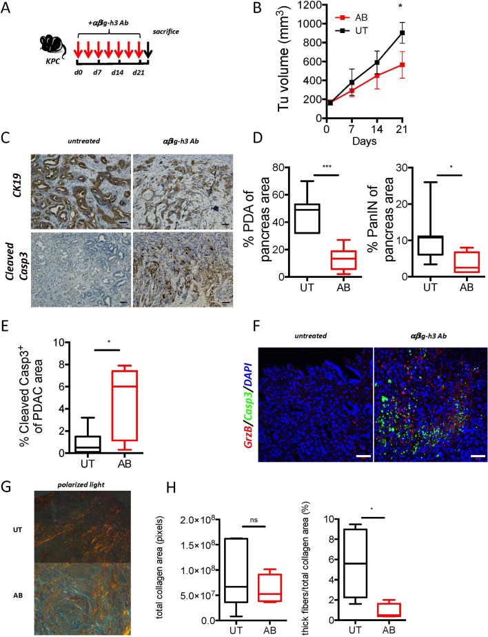
Figure 7.
βig-h3 depletion in established PDA leads to reduced tumour volume. (A) Experimental protocol used for antibody depletion. (B) Tumoural volume was quantified using ultrasound (Vevo2100) in Ab-treated animals. (C) Representative immunohistochemistry for CK19 and cleaved caspase-3 in anti-βig-h3-treated (AB) and untreated (UT) KPC mice. Scale bar, 50 μm. (D) Quantification of PDA and PanIN areas based on CK19 staining and (E) quantification of the results of staining for cleaved caspase-3. (F) Representative immunofluorescence staining for granzyme B, cleaved caspase-3 and DAPI in antiβig-h3-treated and UT KPC mice. Scale bar, 50 μm. The experiment was performed using five to six mice per group. *P<0.05 and ***p<0.001. (H) Quantification of total collagen (transmitted light) and thick fibres (polarised light) content and representative photos in polarised light in treated (AB) and (UT mice. Scale bar 50 μm. *P<0.05, ****p<0.0001. Ab, antibody; KC, p48-Cre; KrasG12D; PDA, pancreatic ductal adenocarcinoma.

