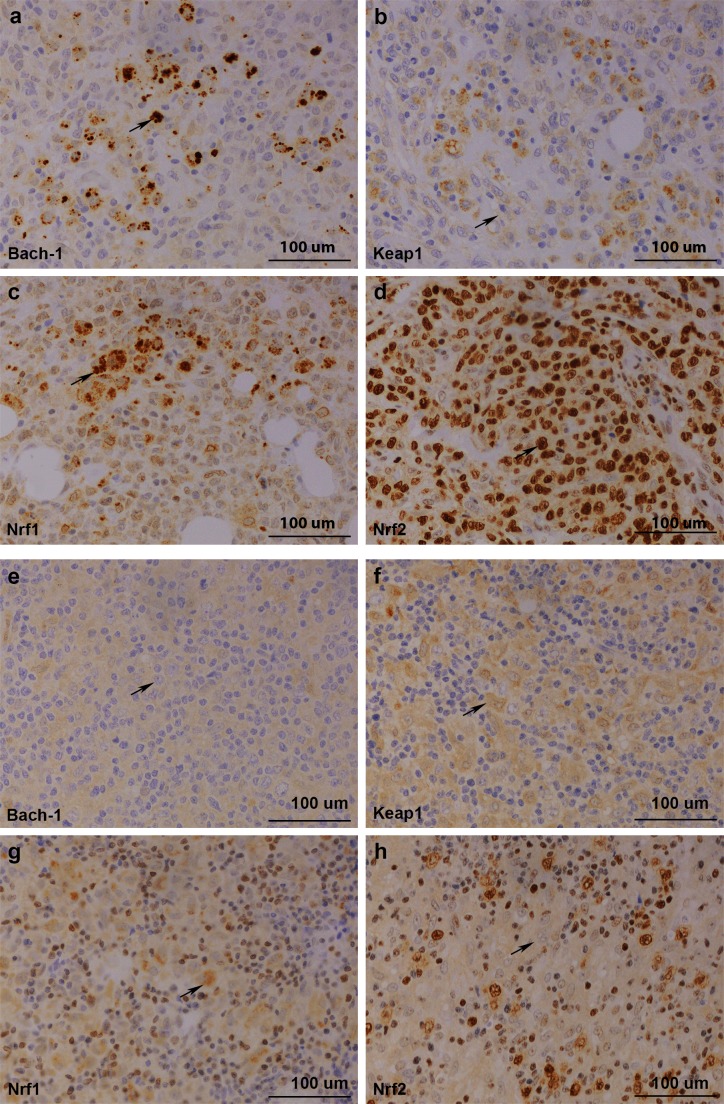
Figure 2.
(A–D) Immunohistochemical staining of patient 1. Biopsy represents non-GC DLBCL from lymph node. From upper left: positive Bach1 expression, low cytoplasmic Keap1 expression, high cytoplasmic Nrf1 expression and high nuclear Nrf1 expression, positive cytoplasmic Nrf2 expression and high nuclear Nrf2 expression. (E–H) Immunohistochemical staining of patient 2. Biopsy represents T cell-rich B-cell lymphoma from lymph node. From upper left: negative Bach1 expression, high cytoplasmic Keap1 expression, high cytoplasmic Nrf1 expression and low nuclear Nrf1 expression, negative cytoplasmic Nrf2 expression and low nuclear Nrf2 expression. GC, germinal centre; Bach1, CNC homolog 1; DLBCL, diffuse large B-cell lymphoma; Keap1, Kelch ECH associating protein 1; Nrf1, nuclear factor erythroid 2-related factor 1; Nrf2, nuclear factor erythroid 2-related factor 2.

