Figure 1.
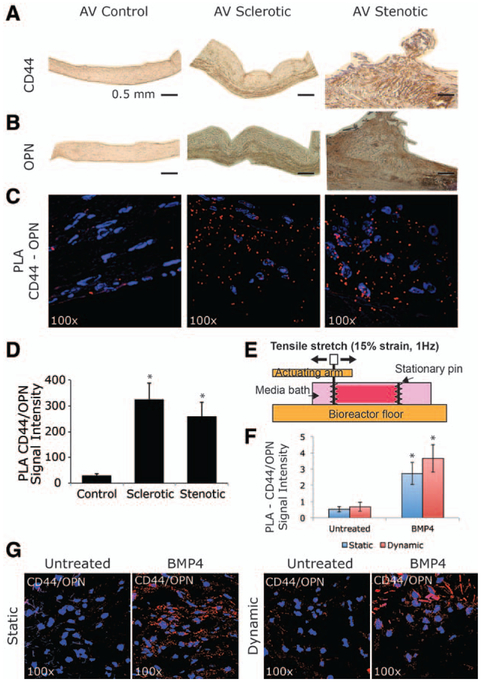
CD44–osteopontin (OPN) functional interaction as a hallmark of early stages of calcific aortic valve disease (A and B). Representative images showing histological analysis of human aortic valve (AV; controls, sclerosis, and stenosis). Immunohistochemistry staining for CD44 and OPN. Bar represents 0.5 mm. C, Proximity ligation assay (PLA), red fluorescent dots indicate reaction product showing extracellular binding between CD44 and OPN. Magnification ×100. D, Bar graph representing PLA quantification in controls, sclerotic, and stenotic aortic valves. *P<0.05. E, Configuration of testing sample in tension bioreactor. F, Bar graph representing PLA quantification in sclerotic aortic valves under static or dynamic (15% stretch at 1 Hz) conditions in the absence or presence of bone morphogenetic protein 4 (BMP4; 100 ng/mL). *P<0.05. G, PLA showing CD44/OPN binding in AV sclerotic tissue at 6 days under static or dynamic (15% stretch at 1 Hz) conditions in the absence or presence of bone morphogenetic protein 4 (BMP4; 100 ng/mL).

