Figure 5.
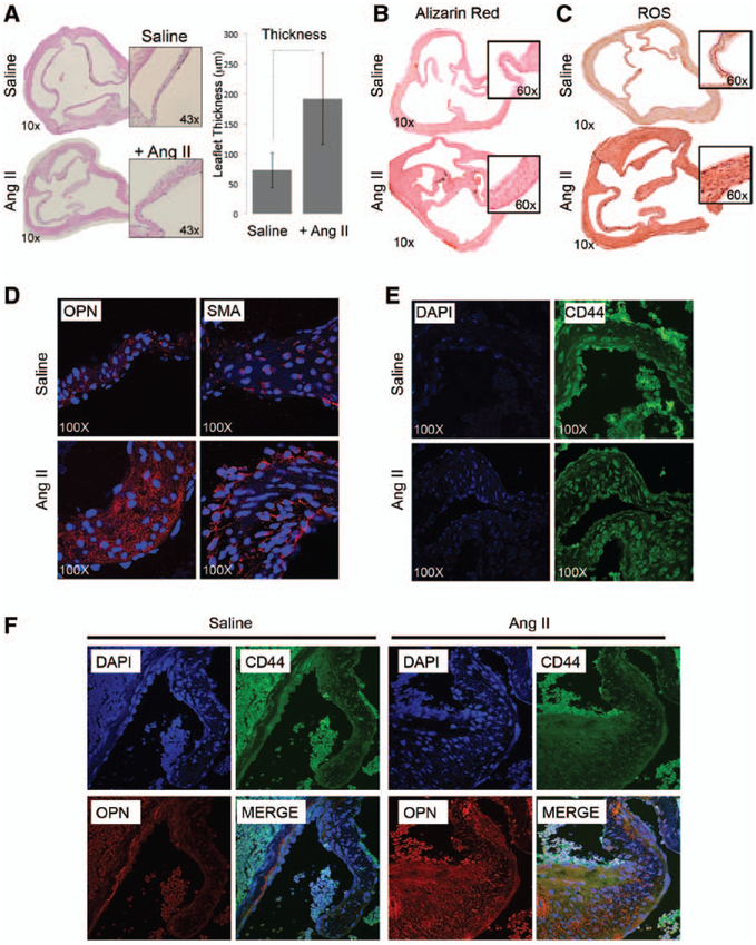
Angiotensin II (Ang II) infusion provokes remodeling of the aortic valve tissue in mice. A, Hematoxylin–eosin staining of cross section of mice hearts harvested 28 days after saline or Ang II infusion (magnification, ×10 and ×43). Bar graph representing leaflet thickness (μm). B, Alizarin red staining showing minimal calcium accumulation in the aortic valve of Ang II-treated mice. C, Nitrotyrosine staining showing oxidative damage accumulation in the aortic valve of Ang II-treated mice (magnification, ×10 and ×60). D and E, Osteopontin (OPN), smooth muscle actin (SMA), and CD44 expression detected by immunofluorescence in aortic valve leaflets. Images were taken using confocal microscopy (magnification, ×100). F, Coimmunofluorescence showing OPN (red) and CD44 (green) colocalization in aortic valve leaflets (magnification, ×60). DAPI indicates 4’,6-diamidino-2-phenylindole; and ROS, reactive oxygen species.

