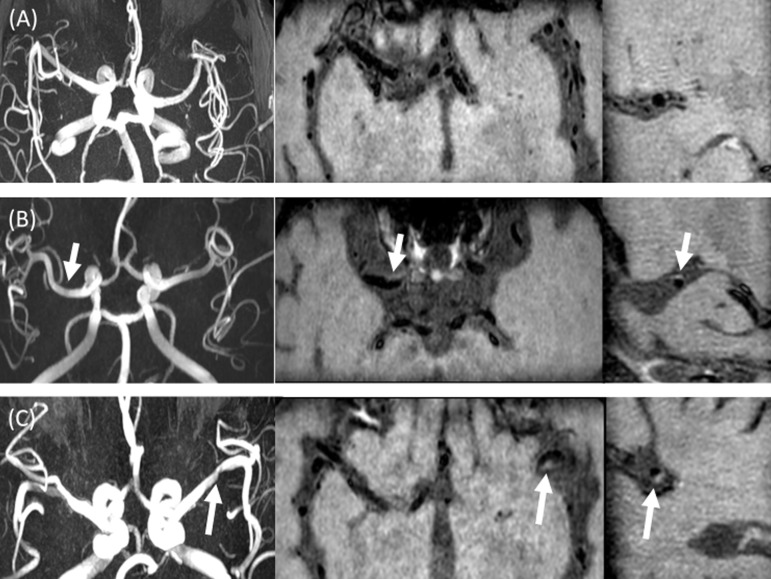
Figure 2.
Examples of plaque classifications. Figure 2A shows an example of “no plaque” category. There is no luminal stenosis on MRA (A, left), without evidence of wall thickening on axial T 1 pre-contrast (A, middle) and sagittal T 1 post-contrast MPR (A, right). Figure 2B shows an example of “possible plaque”. There is no evidence of luminal stenosis on MRA (B, left), with evidence of wall thickening (arrow) on axial T 1 pre-contrast (B, middle) and sagittal T 1 post-contrast MPR (B, right). Figure 2C shows an example of “definite plaque”. On MRA MIP image (C, left), there is focal stenosis of the left M1 MCA (arrow), with corresponding luminal narrowing and wall thickening (arrows) on axial T 1 pre-contrast (C, middle) and sagittal MPR T 1 post-contrast (C, right) images. MCA, middle cerebral artery; MIP, maximum intensity projection; MPR, multiplanar reformat; MRA, MR angiography.

