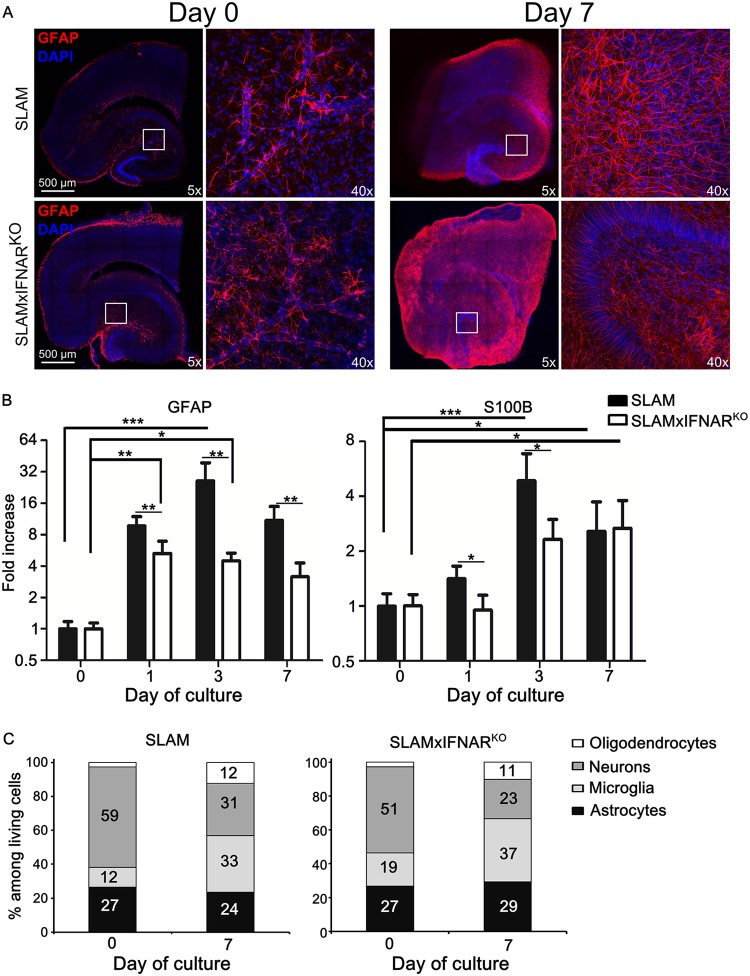
FIG 3.
Astrogliosis and cellular activation during ex vivo hippocampal explant culture. (A) SLAM and SLAM × IFNARKO mouse hippocampus slices (at least 5 slices/animal) were stained with anti-GFAP antibody (an astrocyte marker). Nuclei were counterstained with DAPI. Images were taken at the indicated time of culture with a Leica SP5 confocal microscope and show a high level of activation of astrocyte populations at a low magnification (reconstructed tile; left) or a high magnification (×40 objective; right) in response to slicing and culture procedures. (B) Monitoring of GFAP (an astrocyte proliferation marker) and S100B (a global activation marker) mRNA expression in SLAM and SLAM × IFNARKO mouse hippocampus OBC (n = 5) during the first week of culture by RT-qPCR (normalized to the standard deviation for GAPDH mRNA). The results are presented as the fold increase in expression compared to that on day 0. Statistical analyses were performed using the Kruskal-Wallis test. *, P < 0.05; **, P < 0.01; ***, P < 0.001). (C) The evolution of cell composition over 7 days of culture was studied by flow cytometry both in SLAM and in SLAM × IFNARKO mouse hippocampus OBC (n = 3). The results are presented as the percentage of stained cells among living cells after OBC dissociation.

