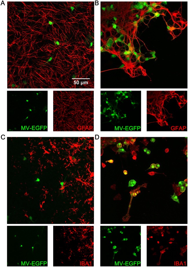
FIG 8.
Confocal analysis of cell susceptibility to MeV-EGFP infection in hippocampus slices at day 7 of culture. (A) Astrocytes in a SLAM transgenic mouse. (B) Astrocytes in a SLAM × IFNARKO transgenic mouse. (C) Microglia in a SLAM transgenic mouse. (D) Microglia in a SLAM × IFNARKO transgenic mouse. Cell staining (i.e., staining for GFAP for astrocytes and IBA1 for microglia) is represented in red, and EGFP is represented in green.

