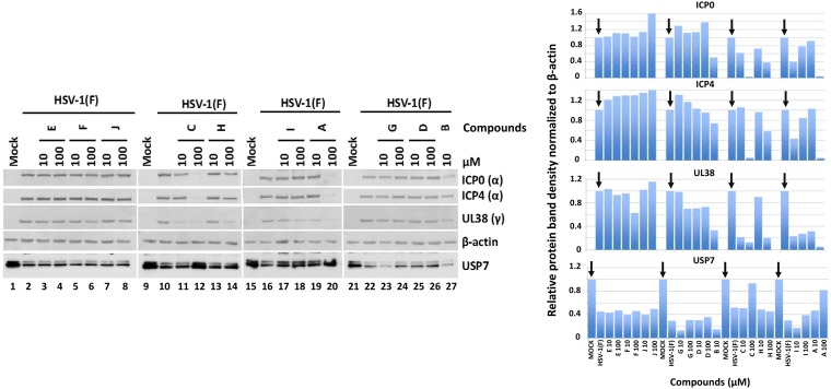
FIG 4.
Effects of compounds on accumulation of ICP0 substrates and on viral gene expression. HEp-2 cells were infected with HSV-1(F) (5 PFU/cell). The compounds were added to the cultures at 10 or 100 μM at the time of infection, and the cells were harvested at 8 h postinfection. Equal amounts of proteins from total cell lysates were analyzed by immunoblot analysis using antibodies against ICP0, ICP4, UL38, β-actin, and USP7 (left). Uninfected cells (lanes 1, 9, 15, and 21) and infected but untreated cells (lanes 2, 10, 16, and 22) served as controls. Quantification of the protein bands was performed using Image J software (right).

