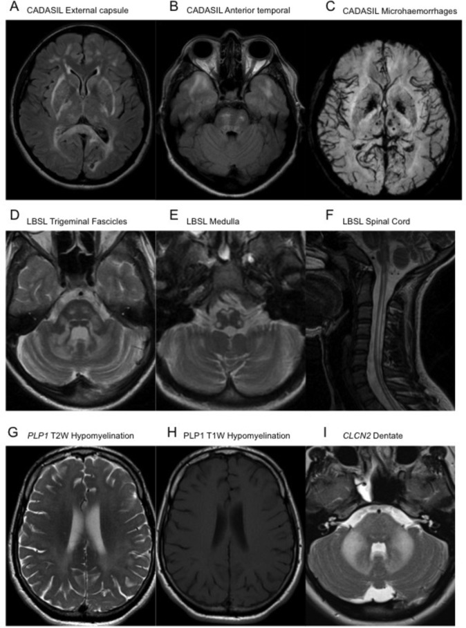
Figure 2.

Axial FLAIR MRI sequences demonstrating confluent signal hyperintensity involving the external capsules, posterior limb of the internal capsules, peritrigonal white matter and splenium of the corpus callosum (A), as well as of the subcortical white matter of the temporal poles and central pons (B) in CADASIL. Minimal intensity projection from a susceptibility weighted imaging acquisition MRI demonstrating multiple punctate foci of paramagnetic susceptibility limited within the thalamus, left putamen and subcortical white matter of the left occipital lobe (C), in keeping with multiple microhaemorrhages in CADASIL. Axial T2W MRI sequences demonstrating signal hyperintensity within the trigeminal fascicles and cerebellar white matter (D), as well as within the pyramids, decussation of the medial lemnisci and inferior cerebellar peduncles at the level of the medulla oblongata (E) in LBSL. A sagittal T2W sequence of the upper spinal cord in the same patient demonstrates contiguous longitudinally extensive signal hyperintensity of the dorsal columns and lateral cortical spinal tracts (F). Axial T2W (G) and T1W (H) MRI sequences in a patient with Pelizaeus-Merzbacher disease illustrating the confluent diffuse T2W hyperintense signal within the white matter of the cerebrum, appearing unremarkable on the T1W sequences, suggestive of hypomyelination. Axial T2W sequences demonstrating confluent hyperintense signal change within the pons, middle cerebellar peduncles and dentate nuclei in a patient with a CNCL2 leukodystrophy (I). CADASIL, cerebral arteriopathy with subcortical infarcts and leukoencephalopathy; FLAIR, fluid-attenuated inversion recovery; T1W, T1-weighted; LBSL, leukoencephalopathy with brainstem and spinal cord involvement with elevated lactate; T2W, T2-weighted.
