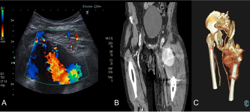
Figure 2:
Diagnosis confirmation by Doppler ultrasound (A), shows hypoechoic lesion with turbulent flow inside with 5–6 cm of maximum diameter; and CT angiography (B) and three-dimensional reconstruction (C) that confirm a lateral circumflex artery pseudoaneurym branch of the deep femoral artery of 7,3 × 6,7 × 6 cm size.

