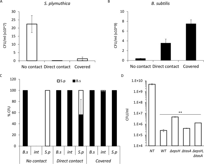
FIG 2.
B. subtilis eliminates S. plymuthica cells. (A) The number of CFU of S. plymuthica isolated from the area of interaction at 24 h (no contact), 48 h (direct contact), and 72 h (covered) after inoculation. Error bars represent standard deviations of the results from 5 biological replicates. (B) CFU counts of B. subtilis isolated from the area of interaction at 24 h (no contact), 48 h (direct contact), and 72 h (covered) after inoculation. Error bars represent standard deviations of the results from 5 biological replicates. (C) Relative CFU counts at each stage of interaction. The interacting colonies were divided into three areas, as follows: B.s, the area of the B. subtilis colony most distant from the interaction area; int, the area of direct interaction; and S.p, the area of the S. plymuthica colony most distant from the interaction area. Each section was separately harvested, sonicated, and plated to determine the number of replicative cells of each species. The experiment was repeated at 24 h (no contact), 48 h (direct contact), and 72 h (covered) after inoculation. (D) Total CFU counts of S. plymuthica following the interaction with wild-type B. subtilis (WT) and the indicated strains. Colonies were inoculated 0.3 cm apart to compensate for the expansion defect. **, P < 0.005 based on a two-tailed Student's t test, compared to S. plymuthica colony grown alone (untreated control [NT]). Error bars represent standard deviations. All experiments were performed at least 3 times with at least three technical repeats.

