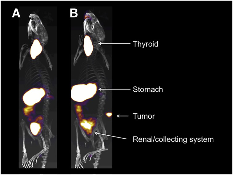
FIGURE 3.
Representative SPECT/CT images of mice with flank prostate-specific membrane antigen–expressing prostate xenografts. SPECT/CT scans were acquired after intravenous injection of 99mTcO4− in animals 16 d after subcutaneous xenograft injection and 9 d after intravenous injection of 1 × 10⁶ hNIS-expressing prostate-specific membrane antigen–targeting CAR T-cells with truncated CAR intracellular signaling domain (A) and fully functional CAR signaling domain (B). Physiologic uptake in thyroid and stomach and clearance of radiotracer via renal collecting system marked by white arrows. In signaling-competent CAR-treated animal, hNIS-expressing CAR T-cells are clearly visualized penetrating flank tumor.

