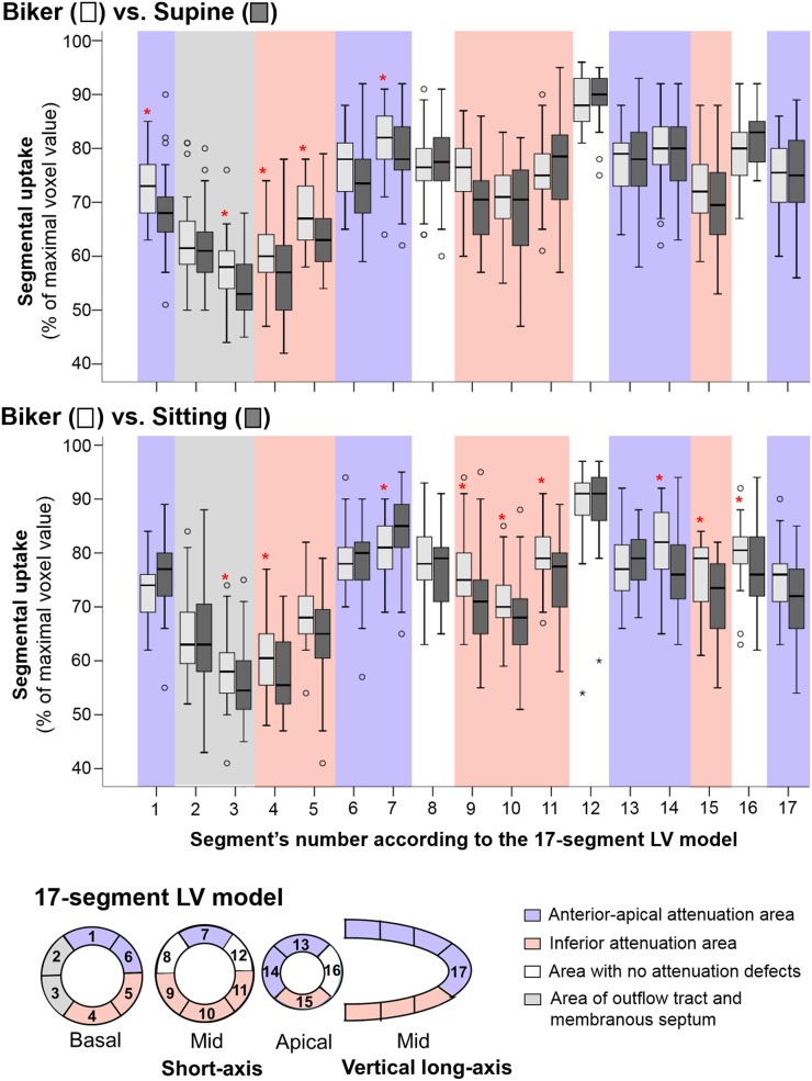
FIGURE 2.
Per-segment distribution of uptake values recorded with biker position, as compared with those obtained in same patients in either supine (top) or sitting (bottom) positions, after exclusion of segments with necrotic or ischemic pattern (with n ranging from 32 to 40 in each segment’s group). Also shown is schematic representation of 17-segment LV model and of inferior and anterior–apical areas where attenuated segments were identified. Segments 2 and 3, corresponding to outflow tract and membranous septum, were excluded from these areas. *P < 0.05 for paired comparisons between patient positions.

