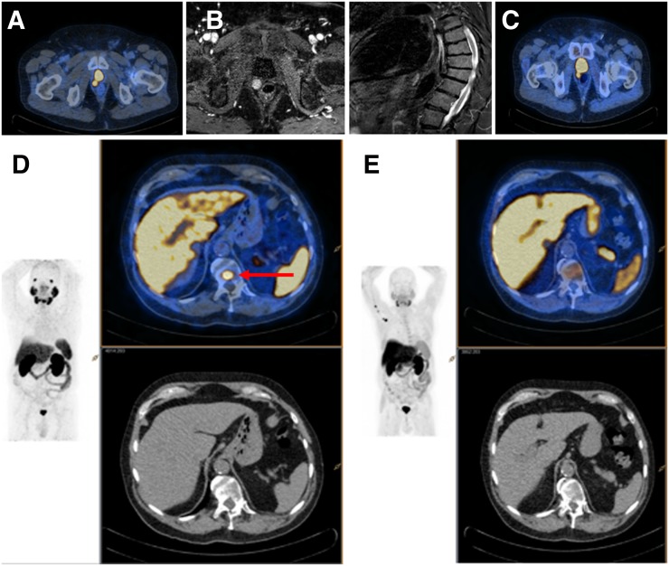
FIGURE 2.
Man with a rising PSA (0.29 ng/mL) 5 y after RP. Imaging demonstrates PF recurrence on PSMA (A), MRI (B), and 18F-FCH (C). Solitary PSMA-avid (D), 18F-FCH (E) and MRI-negative focus in thoracic spine (red arrow) was confirmed as true-positive (repeat imaging and targeted treatment response).

