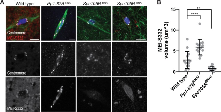
Fig 3. MEI-S332 localizes to centromeres and heterochromatin.
A) MEI-S332 has enhanced recruitment to the pericentromeric regions in Pp1-87B RNAi oocytes and is decreased in Spc105R RNAi oocytes. Two images of Spc105R RNAi oocytes show MEI-S332 localization either abolished or greatly reduced. Confocal images are shown with centromeres (white), MEI-S332 (red), tubulin (green) and DNA (blue). Scale bar indicates as 5 μm. (B) Quantification of MEI-S332 volume. The number of oocytes for measuring are wild type (14), Pp1-87B RNAi oocytes (16) and Spc105R RNAi oocytes (16). Error bars indicate standard deviation, ** = p <0.01 and **** = p <0.0001.

